A Case of Peduncular Hallucinosis Due to Right Pontine and Cerebral Peduncle Cerebrovascular Accident Treated Successfully With Risperidone:
Insights Into an Uncommon Cause of Visual Hallucinations
Peduncular hallucinosis (PH), an uncommon cause of visual hallucinations, is typically due to structural lesions of rostral brainstem or diencephalon.1 We present a case of PH due to right pontine/cerebral peduncle lesion.
Case Report
Our patient was a 69-year-old man who presented to our hospital with left-sided weakness and dysarthria that began 36 hours prior to admission. On admission, the patient’s blood pressure was 161/102 mm Hg, his pulse was 52 bpm, and he was afebrile. Computed tomography of the head showed acute right pontine and cerebral peduncle ischemia. Arterial blood gas, complete metabolic profile, and blood count results were unremarkable. Chest x-ray and urinalysis demonstrated no evidence of infection. The patient began clopidogrel and aspirin for temporary permissive hypertension to slowly correct blood pressure.
The patient’s medical history included hypertension and chronic obstructive pulmonary disease; he was not receiving any medications or oxygen.
The neurologic examination was remarkable for left upper/lower extremity motor strength (0/5) and dysarthric speech. He was noted to have no changes in vision. The remainder of the neurologic examination results were within normal limits, including language, visual fields to confrontation, extraocular movements, and no nystagmus.
Two weeks into admission, the patient reported visual hallucinations, and we were subsequently consulted. The patient described hallucinations consisting of small snakes that appeared recurrently over several minutes during evening/nighttime hours but were not solely hypnagogic/hypnopompic in timing. The patient quickly developed insight into his visual hallucinations. Electroencephalogram demonstrated no evidence of ictal/postictal activity.
He denied depressed mood/anhedonia, delusions, and auditory hallucinations. He was a smoker of 1 pack per day for 40 years but denied alcohol/illicit drug use. Both blood alcohol and urine drug screen results were negative. He was alert and oriented to person, place, and time, with a Mini-Mental State Examination2 score of 27.
We initiated treatment with risperidone 0.5 mg/d taken at bedtime. After 7 days of treatment and at 2-month follow-up, visual hallucinations were not present. Notably, risperidone was discontinued 1 month postdischarge.
Discussion
PH is a form of complex visual hallucinations typically characterized by vivid, formed, well-organized, and non-stereotyped images of people or animals3 involving the whole visual field and occurring over a period of days to several weeks. Most episodes last for a few seconds per minute and generally occur in the evening or in the dark.4 Pathological lesions have been reported in thalamus, midbrain, and pons. PH should be differentiated from other pathological conditions that may be associated with complex visual hallucinations.3
We posit a working diagnosis of PH for our patient. Our rationale includes an acute onset of predominately nocturnal, but not solely hypnopompic/hypnagogic, visual hallucinations occurring proximate to cerebrovascular accident to right pons and cerebral peduncles. His insight into the reality of visual hallucinations is also consistent with PH.5 Finally, while oculomotor abnormalities have been classically described in PH, the literature is currently mixed, and our patient’s oculomotor functions were intact.5 In the absence of visual impairment in our patient, Charles Bonnet syndrome or visual hallucinations associated with visual impairment of any cause—in clear sensorium, with retained insight, and without other psychopathology6—were ruled out, as were neurocognitive disorder, delirium, alcohol use, and illicit medication use.7
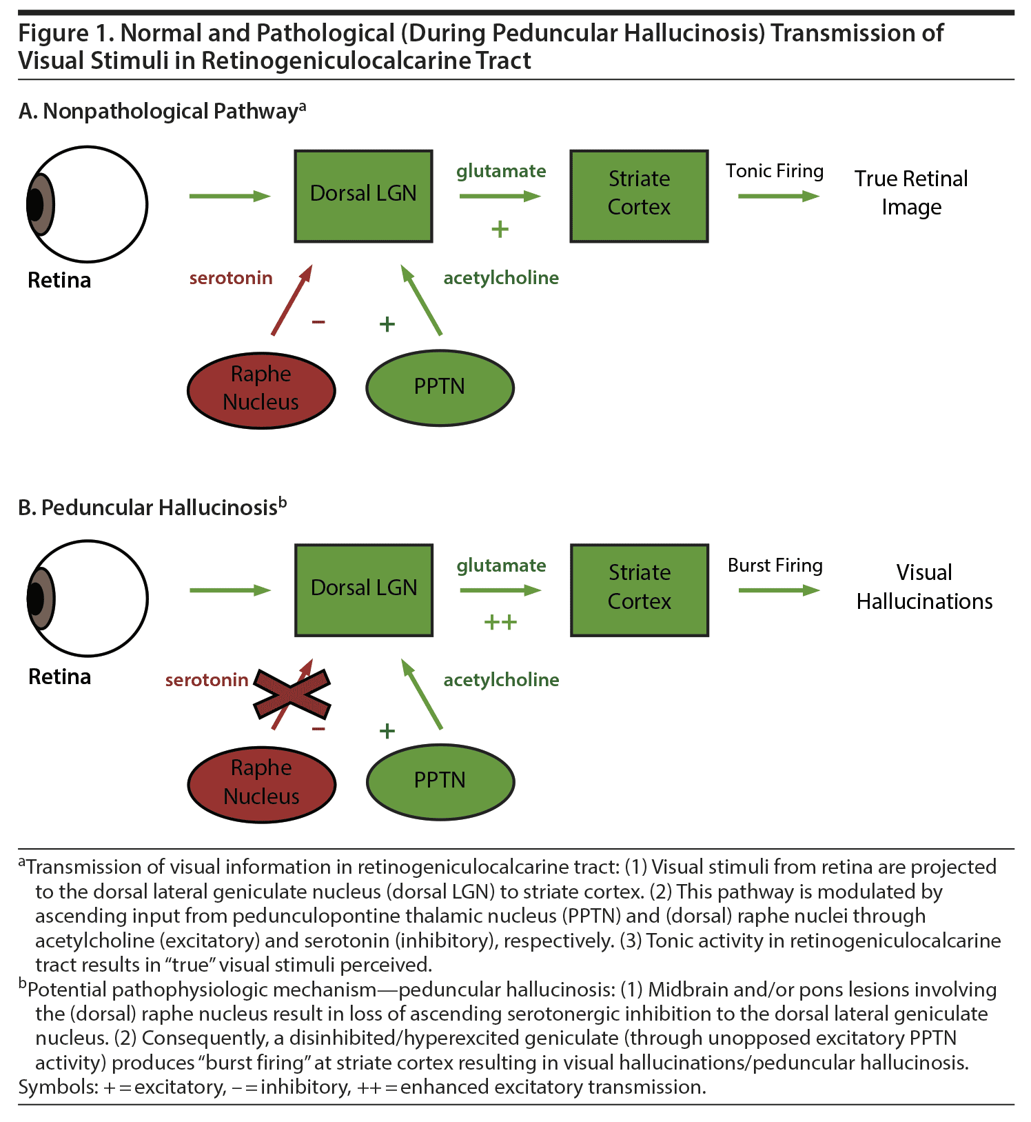
Our proposed pathophysiology of the patient’s visual hallucinations is reviewed in detail in Figure 1.3,5,8
There is a paucity of evidence to guide treatment of PH. It appears that some cases are self-limited. Other studies9-12suggest that atypical antipsychotics could be of potential benefit. In our patient’s case, risperidone was used with resolution of visual hallucinations 7 days afterward and without relapse 2 months after discharge, despite risperidone being discontinued 1 month earlier.
In conclusion, PH, while uncommon, should be considered in the differential diagnosis of visual hallucinations, especially in the elderly or in patients with rostral brainstem and diencephalon lesions.
Published online: October 15, 2020.
Potential conflicts of interest: Dr Spiegel is on speakers bureaus for Allergen, Alkermes, and Otsuka but has no conflict of interest in the preparation of this manuscript. The other authors have no disclaimer/conflict of interest to report.
Funding/support: None.
Patient consent: Permission to publish this case was obtained verbally from the patient, and information about the patient was de-identified to protect anonymity.
REFERENCES
1.Galetta KM, Prasad S. Historical trends in the diagnosis of peduncular hallucinosis. J Neuroophthalmol. 2018;38(4):438-441. PubMed CrossRef
2.Folstein MF, Folstein SE, McHugh PR. "Mini-mental state": a practical method for grading the cognitive state of patients for the clinician. J Psychiatr Res. 1975;12(3):189-198. PubMed CrossRef
3.Notas K, Tegos T, Orologas A. A case of peduncular hallucinosis due to a pontine infarction: a rare complication of coronary angiography. Hippokratia. 2015;19(3):268-269. PubMed
4.Kölmel HW. Peduncular hallucinations. J Neurol. 1991;238(8):457-459. PubMed CrossRef
5.Manford M, Andermann F. Complex visual hallucinations: clinical and neurobiological insights. Brain. 1998;121(pt 10):1819-1840. PubMed CrossRef
6.Winton-Brown TT, Ting A, Mocellin R, et al. Distinguishing neuroimaging features in patients presenting with visual hallucinations. AJNR Am J Neuroradiol. 2016;37(5):774-781. PubMed CrossRef
7.Teeple RC, Caplan JP, Stern TA. Visual hallucinations: differential diagnosis and treatment. Prim Care Companion J Clin Psychiatry. 2009;11(1):26-32. PubMed CrossRef
8.Mocellin R, Walterfang M, Velakoulis D. Neuropsychiatry of complex visual hallucinations. Aust N Z J Psychiatry. 2006;40(9):742-751. PubMed CrossRef
9.Spiegel D, Barber J, Somova M. A potential case of peduncular hallucinosis treated successfully with olanzapine. Clin Schizophr Relat Psychoses. 2011;5(1):50-53. PubMed CrossRef
10.Talih FR. A probable case of peduncular hallucinosis secondary to a cerebral peduncular lesion successfully treated with an atypical antipsychotic. Innov Clin Neurosci. 2013;10(5-6):28-31. PubMed
11.Spiegel DR, Lybeck B, Angeles V. A possible case of peduncular hallucinosis in a patient with Parkinson’s disease successfully treated with quetiapine. J Neuropsychiatry Clin Neurosci. 2009;21(2):225-226. PubMed CrossRef
12.Vetrugno R, Vella A, Mascalchi M, et al. Peduncular hallucinosis: a polysomnographic and SPECT study of a patient and efficacy of serotonergic therapy. Sleep Med. 2009;10(10):1158-1160. PubMed CrossRef
aDepartment of Psychiatry and Behavioral Sciences, Eastern Virginia Medical School, Norfolk, Virginia
*Corresponding author: David R. Spiegel, MD, Eastern Virginia Medical School, Department of Psychiatry and Behavior Sciences, 825 Fairfax Ave, Norfolk, VA 23507 ([email protected]).
Prim Care Companion CNS Disord 2020;22(5):19l02584
To cite: Spiegel DR, Harsch C, Pattison A. A case of peduncular hallucinosis due to right pontine and cerebral peduncle cerebrovascular accident treated successfully with risperidone: insights into an uncommon cause of visual hallucinations. Prim Care Companion CNS Disord. 2020;22(5):19l02584.
To share: https://doi.org/10.4088/PCC.19l02584
© Copyright 2020 Physicians Postgraduate Press, Inc.
Please sign in or purchase this PDF for $40.00.
Save
Cite

